The National Institute of Health document linked below outlines the equipment used at the MRRF, and provides useful information for researchers:
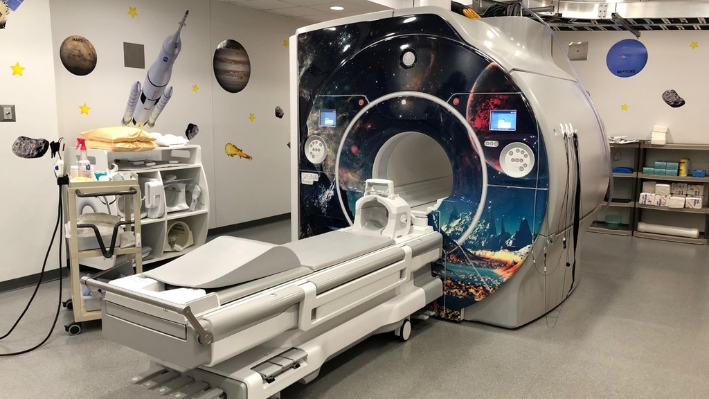
3.0T GE SIGNA Premier MRI Scanner
The research-dedicated 3.0T GE SIGNA Premier MRI scanner is located in L425 PBDB (Pappajohn Biomedical Discovery Building) and is available from 8:00am to 6:00pm Monday through Friday.
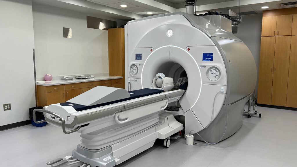
GE MAGNUS 3T Head Only Scanner
The research-dedicated 3.0T GE MAGNUS MRI scanner is located in L425 PBDB (Pappajohn Biomedical Discovery Building) and is available from 8:00am to 6:00pm Monday through Friday.
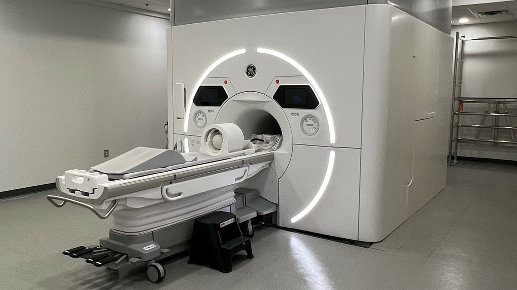
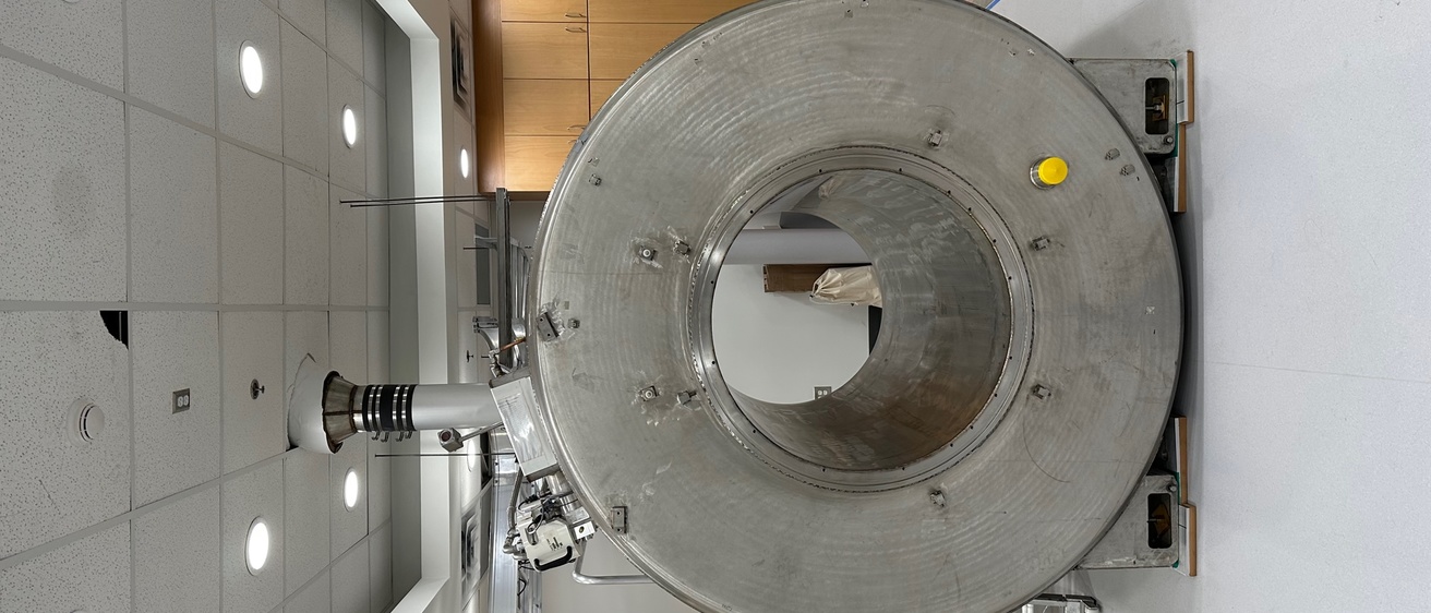
GE Signa 7T Whole Body Scanner
The research-dedicated GE Signa 7T Whole Body Scanner is located in L550 PBDB (Pappajohn Biomedical Discovery Building) and is available from 8:00am to 5:00pm Monday through Friday.
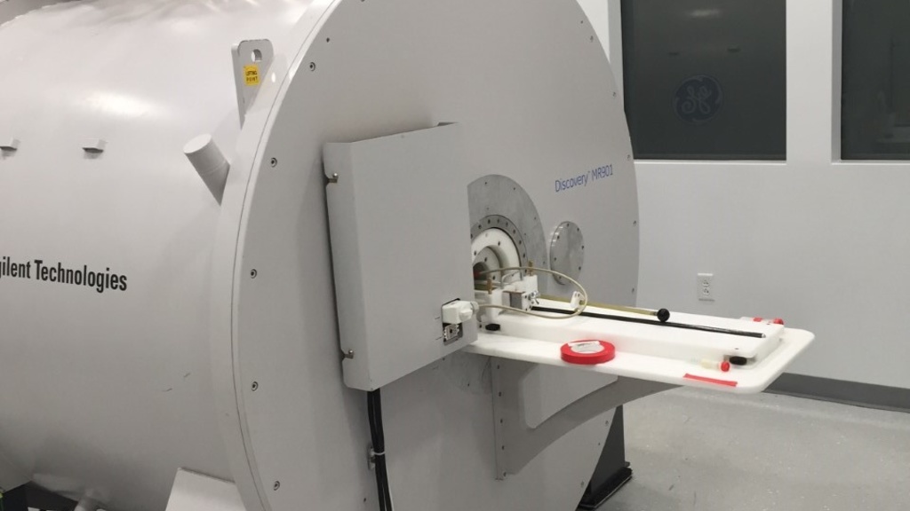
7.0T GE 901 Discovery MRI Small Animal Scanner
The 7.0T GE 901 Discovery MRI Small Animal Scanner is located in the PBDB (Pappajohn Biomedical Discovery Building) and is available from 8:00am to 5:00pm Monday through Friday.
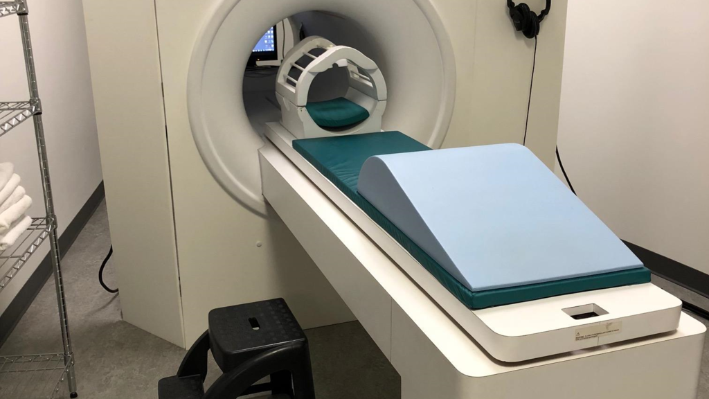
MRI Simulator
The MRI Research Facility's MRI Simulator is available to all MRI researchers at no charge. The MRI Simulator gives subjects exposure to the scanner by realistically reproducing the scanning environment.
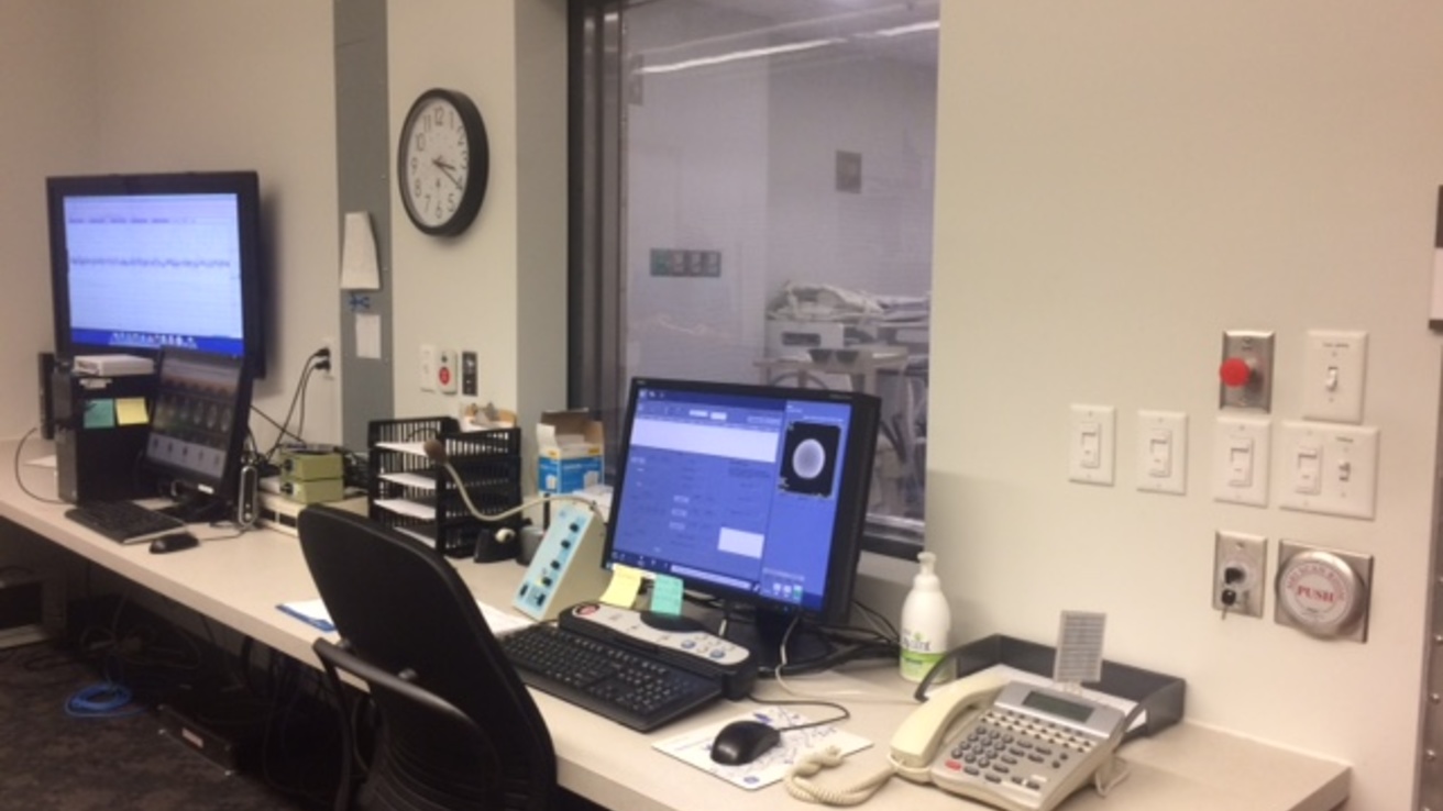
Computing Facilities
Within the MRRF Imaging Facility, research workstations are available for investigators to utilize while imaging and post processing data after it is collected. Both Microsoft Windows and Linux-based workstations are available. Several image processing tools are available on these systems including:
- BRAINS
- Slicer
- FSL
- AFNI
- MatLab
- LCModel
We are actively involved with the Iowa Institute of Biomedical Imaging (IIBI) and the Institute for Clinical and Translational Science (ICTS) technical groups to provide administration of our computing resources. IIBI provides system upgrades, on-site technical support and backups for all research machines. ICTS currently provides support for the MRI Research Facility's website, scheduling system and the LCModel server.
The MR Research Facility uses the XNAT (Extensible Neuroimaging Archive Toolkit) open source software platform to manage all MR research images. XNAT allows researchers to access and manage their MR images from a secure repository immediately following a subject's research scan. Subject data is confidential and is available for all current MR research projects.
Other Equipment
The MR Research Facility strives to keep our equipment state-of-the-art and to provide tools to investigators that will facilitate their research projects. Below is some of the equipment we provide to all MR Research Facility users:
Other Equipment
BIOPAC Physiological Monitoring System
Includes Photoplethysmograph (PPG), Respiratory, Galvanic Skin Response (GSR), Pulse oximeter, air flow and expired gas analysis.
fMRI MediGoggles
MediGoggles permit subjects who require prescription glasses to see visual stimuli from within the scanner without introducing any safety risks to the subject or artifact in the images. The goggles can be customized from –6 to +6 dioptre in 0.5 dioptre increments and are easy to set up and use.
FOMRI II Dual Channel MRI Microphone System
The FOMRI II system uses fiber-optic technology and a complex set of adaptive acoustic algorithms to achieve high levels of noise reduction with no loss of speech quality.
Metrasens FerroGuard Freestanding Metal Detector
The Metrasens FerroGuard metal detector is available at the research suite at L400 PBDB and is used to screen every research subject for potential hazardous metallic material. The system is movable and can be adjusted to meet the sensitivity requirements of the location.
Avotec SV-6060 HDMI LCD MRI Scan Room Projector
The SV-6060 projector system is an HDMI resolution, LCD Projector specifically designed to be used in the MR magnet room.
- Entertainment / Research
- Can be located anywhere in the magnet room. No extra wave guides or mirrors required.
The Avotec SV-6060 video projection system is installed at the following MRI Scanners:
- GE 3T Premier full body
- GE 3T MAGNUS head only
- GE 7T full body
Current Designs Fiber Optic Response Systems
The Current Designs 932 interface with Pyka 8 Button Bimanual (pinkies disabled) handheld response device systems have been installed at the following MRI scanners:
- 3T Premier full body
- 3T MAGNUS head only
- 7T full body scanner
- Simulator
The 932 opto-electronic interface receives optical signals from the handheld devices in the MR suite and converts them into electronic signals for the computer.
The outputs are not designed to be used with a specific program or hardware platform but are instead designed to work with everything. This interface provides complete USB, Serial and Parallel outputs.